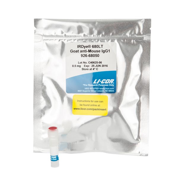
Highly cross-adsorbed goat anti-mouse IgG1 (γ 1 chain specific) antibody conjugated to IRDye 680LT.
Immunogen
Mouse IgG1 paraproteins
Purity and Specificity
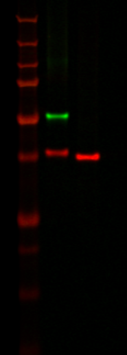
Isolation of specific antibodies was accomplished by affinity chromatography on mouse IgG1 covalently linked to agarose. Based on ELISA and flow cytometry, the antibody reacts with the heavy chain of mouse IgG1. This antibody has been tested by dot blot and/or solid-phase adsorbed to ensure minimal cross-reactivity with mouse IgM, IgG2a, IgG2b, IgG3, and IgA, pooled human sera, and purified human paraproteins. The conjugate has been specifically tested and qualified for Western blot applications. Results with your primary antibody may vary and specificity should be confirmed prior to performing two-color detection.
Applications
Recommended for:
- Western Blot
- Protein Array
- Immunohistochemistry
- Microscopy
- 2D Gel Detection
- Tissue Section Imaging
Not Recommended for:
- Small Animal Imaging
- In-Cell Western Assay
- On-Cell Western Assay
Formulation
IRDye 680LT Secondary Antibodies are supplied as purified immunoglobulin conjugates, lyophilized in phosphate-buffered saline, pH 7.4. Protect from light. Store at 4 °C prior to reconstitution.
Each vial contains 10 mg/mL BSA (free of IgG and protease) as a stabilizer and 0.01% sodium azide as a preservative, after reconstitution. Concentration is 1.0 mg/mL when reconstituted as directed. Refer to the pack insert for details on reconstitution.
Recommended Dilutions
| Application | Recommended | Suggested Range |
|---|---|---|
| Odyssey Western blot detection | 1:20,000 | 1:10,000 - 1:50,000 |
| Other | User optimized |
Optimum dilutions will vary and should be determined empirically.
RRID
- P/N 926-68050: RRID AB_2783642
Example Data

