X-ray film can introduce various issues into your Western blot, from requiring several exposures and darkroom expenses to losing strong and faint signals. Digital imaging helps reduce this variability and provide better data reproducibility and confidence in your results. With digital imaging, you can capture more data in a single exposure and utilize a broader dynamic range, which helps you generate consistent, credible results.
Capture Data in a Single Exposure
With X-ray film, several exposures of differing durations are usually required to capture your desired image. Exposure times must be long enough to detect the desired target but short enough to limit undesired background. Additionally, expensive darkroom maintenance, film, and developing reagents are required for imaging your membranes.
Digital imaging can help solve these issues. The Odyssey® XF, for example, minimizes the need for multiple images by capturing your data in a single acquisition. Digital imaging consequently eliminates any need for darkrooms as well as the expenses of film and developing reagents.
Increase Data Accuracy with Near-Infrared Fluorescence
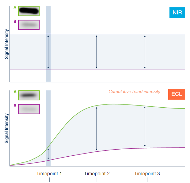
Chemiluminescence uses enzymatic reactions to detect proteins; therefore, it can be challenging to capture these reactions for each target and to successfully quantify the amount of protein on the membrane. Digital imaging with near-infrared (NIR) fluorescence has a more stable signal that can sidestep these issues and help you quantify your data easily.
Stable fluorescent signals deliver consistent, proportional results for quantitative analysis. Relative comparison of band intensity is based on consistent, reproducible signal output. With NIR fluorescence, relative band intensity is constant and proportional across time, every time. But with chemiluminescent detection, the enzymatic reaction is constantly changing. You will see different band intensities at different times and get different answers. These time-dependent changes reflect the limitations of chemiluminescence, not real changes in your samples.
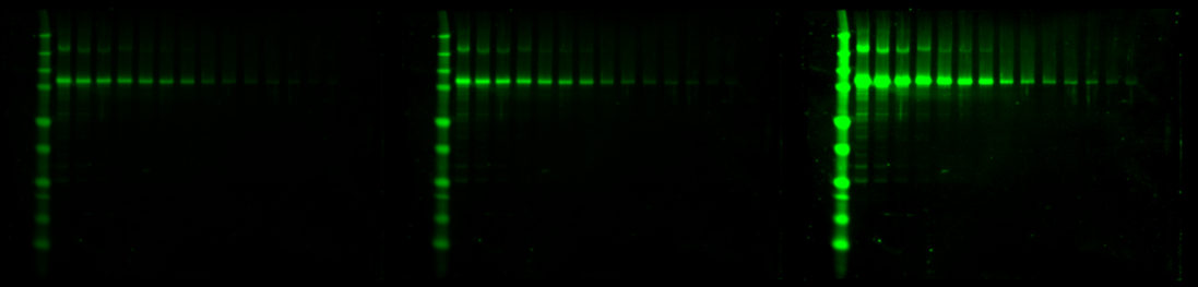
An imager like the Odyssey XF is useful for both chemiluminescence and NIR fluorescence research. With the largest dynamic range of any available digital imager, it can capture the strongest and faintest bands in a single image without saturation.
X-ray film can cause many unnecessary complications for you and your research. Digital imaging helps to mitigate these issues. It captures more data in a single image, does not need a darkroom or film expenses, and captures more bands with a wider dynamic range. Switch to digital imaging systems for help in generating more reliable, consistent results.
We encourage you to check out the Odyssey XF or Odyssey M Imaging System for yourself to learn how you can get more accurate digital images of your data. If you still have questions or concerns about digital imaging, then we hope you'll contact us today for more information. Our team is always eager to help you achieve the best results possible.
Powered by Froala Editor