Enhanced chemiluminescence (ECL) or chemiluminescent Western detection is a traditional method for conducting Western blotting research. A distinguishing characteristic is the use of an enzymatic reaction for protein detection.
Chemiluminescent Western detection begins when the primary antibody first recognizes the target protein or antigen on the membrane. The secondary antibody, which is labeled with an enzyme, binds to the primary antibody. For its enzyme, ECL typically uses horseradish peroxidase (HRP) or alkaline phosphatase (AP). The blot is then incubated with substrate to generate a signal, or a production of light. The most common chemiluminescent detection substrate, HRP, is luminol-based. Consequently, HRP causes the substrate to oxidize and transiently produce a signal in the presence of hydrogen peroxide (H2O2). The light photons are then measured with one of two detection methods: x-ray film or digital imaging.
While chemiluminescent detection is widely used, there are certain aspects to keep in mind when considering it as a Western blotting method.
Ultimately, it can be difficult to capture the enzymatic reaction for each target simultaneously and at the same point each time. This makes it challenging to accurately quantify the protein abundance on the membrane. Additionally, chemiluminescence is unable to multiplex signals or simultaneously detect two targets with the same molecular weight in a single sample that is on the same blot. Overall, these factors make it difficult to achieve precise quantification. It is therefore important to understand the variability of ECL in its entirety before proceeding with experimentation.

The use of x-ray film is a common practice in Western blot imaging. It is essential to know not only the way it works but also how several pertinent factors may introduce variability and error into the data.
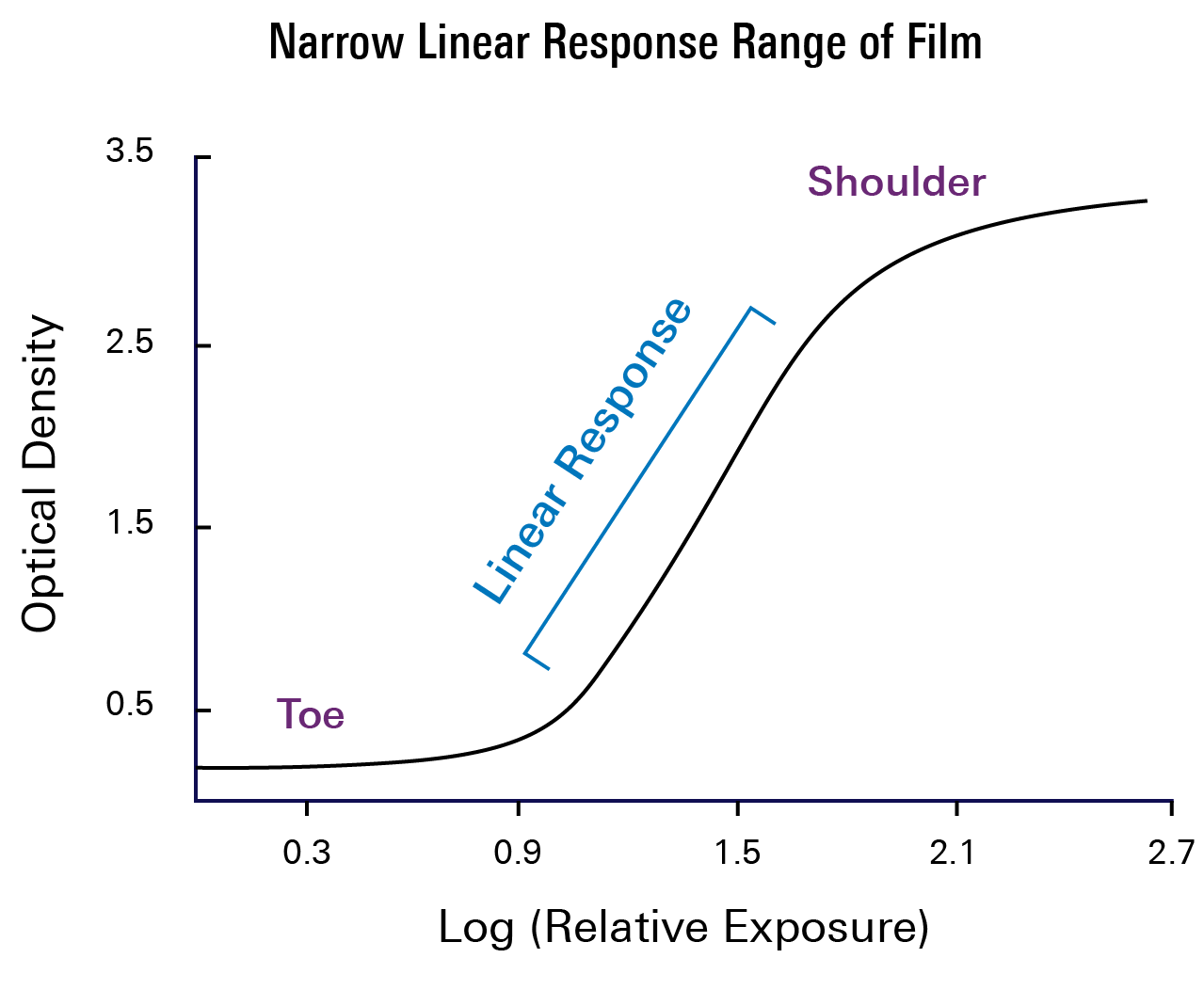
X-ray film is coated with an emulsion that contains light-sensitive silver halide crystals, or “silver grains.” When silver atoms in a grain are light-activated, they reduce the entire grain to black metallic silver. This produces a darkened spot on the x-ray film, or the visual product. Grains that were not exposed to light are washed away.
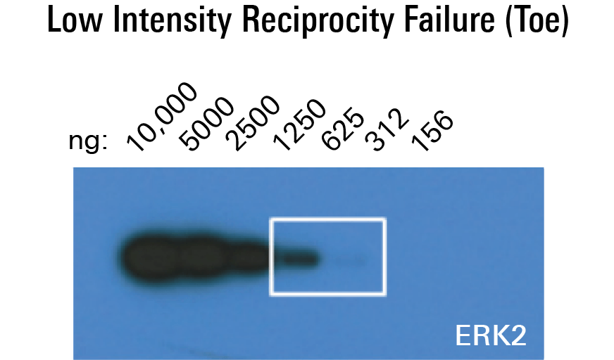
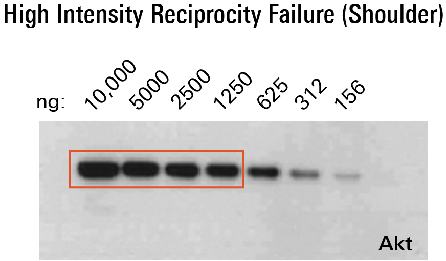
Consequently, x-ray film is effective at confirming the presence or absence of a signal. However, there are several factors to consider before using x-ray film to image a blot. For instance, film has a limited dynamic range, and signals outside that range can go undetected. Faint bands can also go undetected by film, and strong signals can cause film to saturate. Saturated bands can become comparably dark in appearance and make it difficult to distinguish individual bands. Because the protein amount on the membrane is therefore underrepresented by band intensity, this can prevent accurate quantification as well.
Finally, an initial number of photons are required to convert the silver grains in the film; photons are therefore being produced without generating a black signal. This commonly leads to an underrepresentation before the linear response and the shoulder.
LI-COR can help bypass any potential x-ray film constraints by facilitating a transition to digital imaging. Digital imaging is useful because it captures more data than x-ray film in a single exposure and reduces the need for multiple exposures. Additionally, its wider dynamic range can collect more data and therefore decrease the loss of very strong and weak bands.
Depending on the nature of an experiment, there are various digital imaging options that can help generate consistent and credible results. For chemiluminescent research, the C-DiGit® Blot Scanner is useful because it eliminates the need for a darkroom along with film and developing reagents expenses. With a larger dynamic range than film, the C-DiGit scanner can capture more data in an acquisition and minimizes the need for multiple exposures.
For a digital imager capable of both chemiluminescence and near-infrared fluorescence (NIR), utilize the Odyssey® XF. With the largest dynamic range of any available chemiluminescence digital imager, it captures the strongest and faintest bands in a single image without saturation. Unlike other imagers, the Odyssey XF does not introduce image manipulation, such as binning and stacking, during image acquisition. It can facilitate data analysis not only for an individual experiment but between researchers as well.
LI-COR can help mitigate x-ray issues with digital imaging. The C-DiGit scanner, for example, is a practical and useful option for chemiluminescent research. However, the limitations of enzymatic detection are inherent to chemiluminescence. To eliminate these factors completely, consider transitioning to NIR fluorescence with imagers like the Odyssey XF, which can still use current chemiluminescence protocols. When switching to NIR fluorescence entirely, imagers like the Odyssey DLx have the complete capacity of NIR imaging and can be versatile tools for imaging not only Western blots but plate-based assays, slides, and excised organs as well.
Check out the Advanced Applications Brochure for additional information.
There are many other important aspects to chemiluminescence, x-ray film, and digital imaging. Want to learn more? Contact LI-COR today and let us help choose the right detection and imaging methods for you.
See some published examples of Western Blots
View publications