IRDye® 800CW Rabbit anti-HRP
Components (926-32202)
IRDye 800CW Rabbit anti-HRP, 0.5 mg
Applications
IRDye 800CW Rabbit anti-HRP provides a way to convert chemiluminescent Western blots to the near-infrared fluorescence. Incubation of Western blots with IRDye 800CW Rabbit anti-HRP enables detection of HRP-conjugated antibody.
Specifications
Antibody Concentration: 1.0 mg/mL when reconstituted as directed
Fluorophore: IRDye 800CW
Excitation wavelength: 778 nm (in PBS)
Emission wavelength: 795 nm (in PBS)
Form of Antibody: IRDye 800CW conjugated purified immunoglobulin, lyophilized in phosphate buffered saline, pH 7.4. Contains 10 mg/mL BSA (IgG and protease free) as stabilizer and 0.01% sodium azide as preservative.
Contains sodium azide.
Immunogen: Peroxidase from horseradish roots
- Membrane Types: Nitrocellulose or PVDF
Storage: 4 °C
Purity and Specificity
The antibody was purified from antisera by immunoaffinity chromatography using antigens coupled to agarose beads. Based on immunoelectrophoresis and/or ELISA, the antibody reacts with peroxidase from horseradish roots. It may cross-react with peroxidase from other sources. The conjugate has been specifically tested and qualified for Western blot applications.
Reconstitute Secondary Antibody
Combine the contents of vial with 0.5 mL of sterile distilled water.
Gently mix by inverting.
Allow the solution to stand at room temperature for at least 30 minutes before use.
If the solution is not completely transparent after standing at room temperature, centrifuge the solution.
Storing Reconstituted Secondary Antibody
- Reconstituted secondary antibody is stable for up to 3 months at 4 °C when stored undiluted and protected from light.
- Only dilute the reconstituted antibody immediately before use.
Prepare Diluent
This procedure will yield diluent of a final concentration of approximately 0.2% Tween® 20. Alternatively, premade diluents that already contain Tween 20 (i.e. or ) can be used to eliminate this manual step from the procedure.
Add 1.0 mL of 20% Tween 20 to a 100 mL bottle of or .
If you are using a PVDF membrane, add SDS to a final concentration of 0.002 to 0.01% to the antibody diluent.
Mix well by inverting.
Protocol
A Western blot that has been incubated with an HRP conjugated antibody can be detected with IRDye 800CW anti-HRP. Optimal results are obtained when using a Western blot that has not been processed with a chemiluminescent substrate.
Notes
- Do not write on membranes with regular ink pens or markers, because the ink will fluoresce on Odyssey Imaging Systems. You can write on nitrocellulose membranes with pencil or the Odyssey Pen (PN 926-71804). Use only a pencil to write on PVDF membranes, because the ink from the Odyssey Pen will dissolve in the methanol used to wet the PVDF membrane.
Odyssey Blocking Buffer (discontinued) contains 0.1% sodium azide, so it should not be used as an antibody diluent for HRP-conjugated antibodies.
Steps
Dilute IRDye 800CW anti-HRP 1:1000 in antibody diluent (see Prepare Diluent).
Add diluted IRDye 800CW Rabbit anti-HRP conjugate to blot and incubate for 1 hour at room temperature on platform shaker, protected from light.
Decant diluted conjugate and rinse blot briefly in 15 mL of 1X PBST. Decant. Add 15 mL of 1X PBST and incubate at room temperature on platform shaker protected from light for 5 minutes. Decant. Repeat wash step 2 more times.
Rinse with 15 mL of 1X PBS.
Scan wet on an Odyssey Imaging System using the Membrane preset with the 800 nm Channel. Start with Scan Intensity set at 6 and adjust as needed.
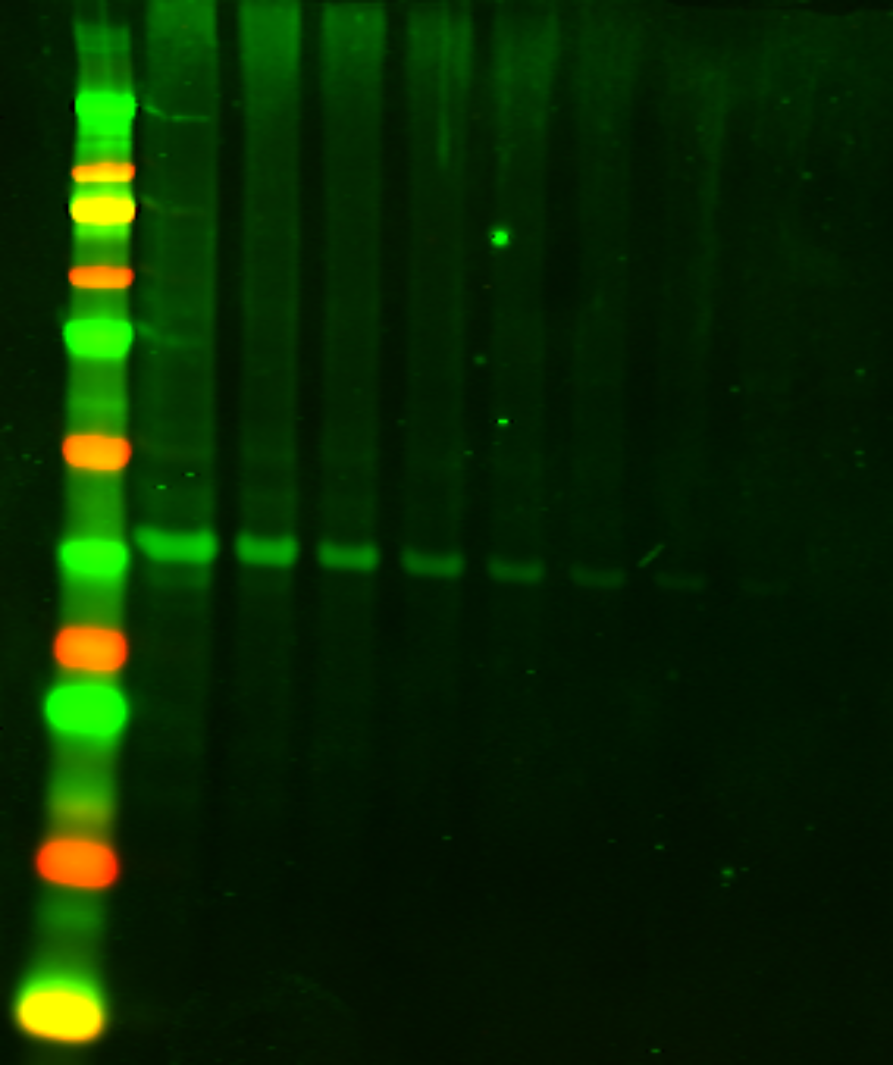
Example Data Using Intercept (PBS) Blocking Buffer (927-60001)

Example Data Using Intercept (PBS) Protein-Free Blocking Buffer (927-80001)
