IRDye® 800CW 2-DG Optical Probe
IRDye® 800CW EGF Optical Probe
IRDye® 800CW RGD Optical Probe
IRDye® 800CW BoneTag™ Optical Probe
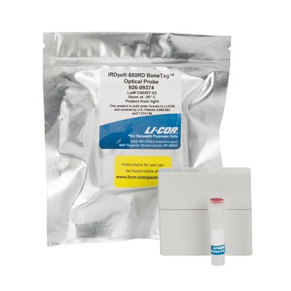
IRDye® 680RD BoneTag™ Optical Probe
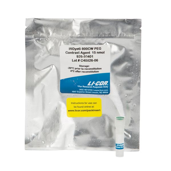
IRDye® 800CW PEG Fluorescent Contrast Agent
Every LI-COR BrightSite IRDye in vivo imaging agent has been carefully validated with cultured cell assays, microscopy, in vivo imaging of animal models, and histology to ensure high affinity and specificity.
BrightSite IRDye Small Animal Imaging Agents are:
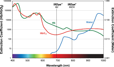
IRDye 800CW absorption/emission near 800 nm matches near-infrared absorption minima for bodily fluids and tissues, resulting in excellent tissue penetration, making it ideal for in vivo imaging.
CellVue® fluorescent imaging kits use proprietary labeling technology to stably incorporate fluorescent dyes containing long aliphatic hydrocarbon tails into lipid membranes.1 They are useful for researchers working in all aspects of science and technology where fluorescently-labeled cells and/or tissues are required. CellVue dyes also provide researchers with valuable tools for many in vivo and in vitro cell studies using fluorescent membrane labels.
CellVue dyes consist of long aliphatic hydrocarbon tails linked to a polar fluorescent chromophore.
| Application | IRDye EGF | IRDye RGD | IRDye 2-DG | IRDye PEG | IRDye BoneTag | CellVue |
|---|---|---|---|---|---|---|
| Tumor Imaging | ||||||
| Metabolic Imaging | ||||||
| Inflammation/Arthritis | ||||||
| Vasculature (Contrast) | ||||||
| Lymphatic Imaging | ||||||
| Lymph Node Imaging | ||||||
| Structural Imaging | ||||||
| Cell Trafficking |